40 how to label gel electrophoresis images
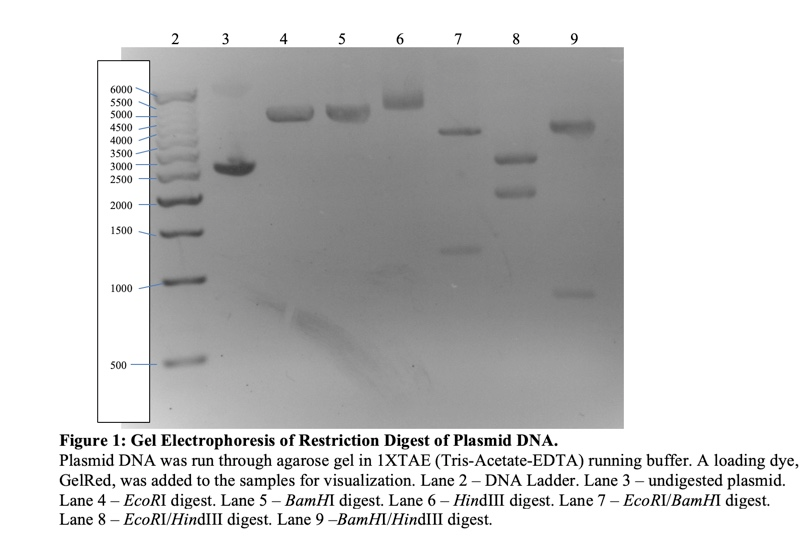
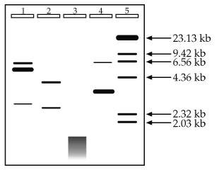
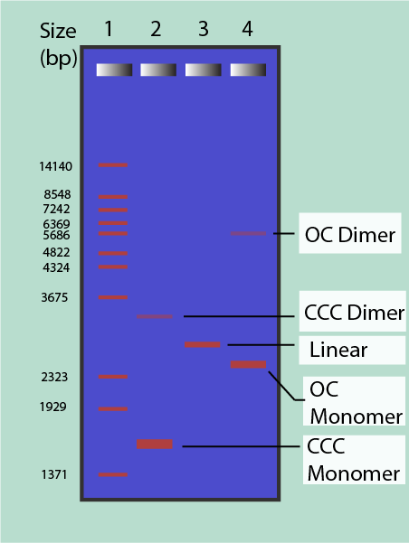
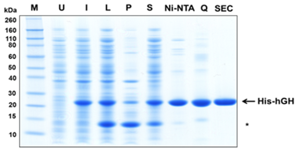
How to Interpret DNA Gel Electrophoresis Results | GoldBio Gel Electrophoresis. Lane 1: DNA Ladder. Lane 2: Undigested plasmid A. Lane 3: Completely digested plasmid A. Lane 4: Digested PCR product (or DNA Fragment). Lane 5: PCR Product (with a faint primer dimer band). Lane 6: Genomic DNA. The white arrows indicate the bands that you want to excise. Gel Electrophoresis - Everything You Need To Know - The Lab Label Step 1: Making and agarose gel. Step 2: Setting up the power box. Step 3: Loading the samples and the molecular weight ladder. Step 4: Running the gel. Step 5: Staining the gel and analysing the gel. Tips and tricks to get the best gel electrophoresis results. The preperation and concentration of the agarose gel.
Publication-quality labeling of gels using transparency-mounted ... Obtaining a high quality nucleic acid or protein gel may be an easier and less daunting task than labeling the gel for publication. Techniques for adding information and identifiers to gels range from laboriously applying lettering or labels on gel photographs to computerized approaches that work directly with digitized gel images and produce photographic-quality output.

How to label gel electrophoresis images
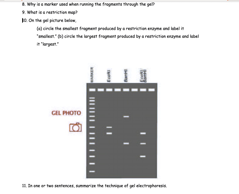
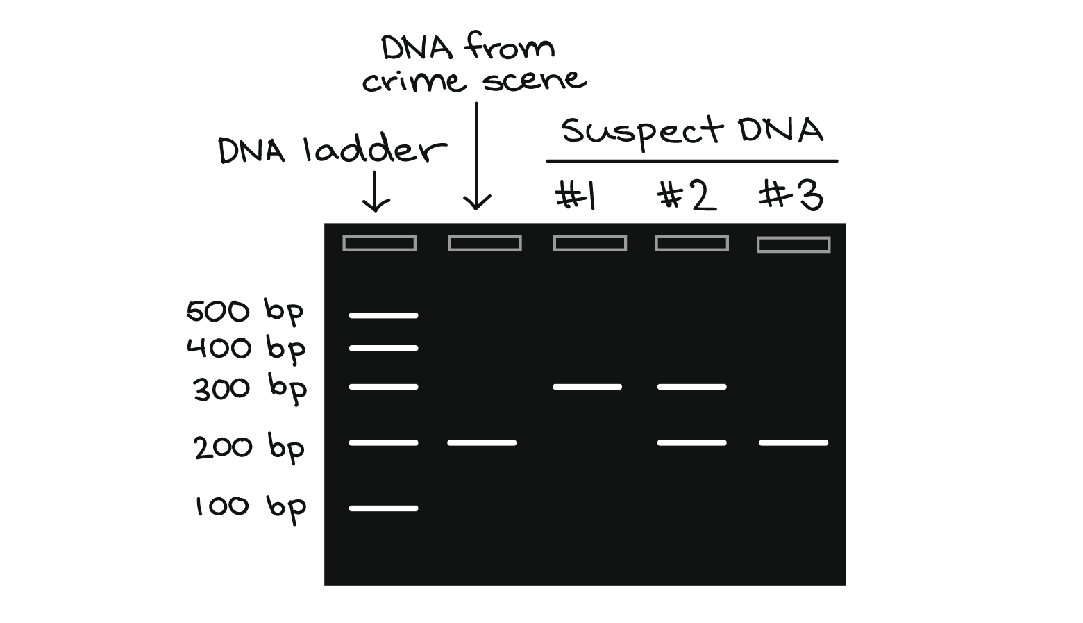
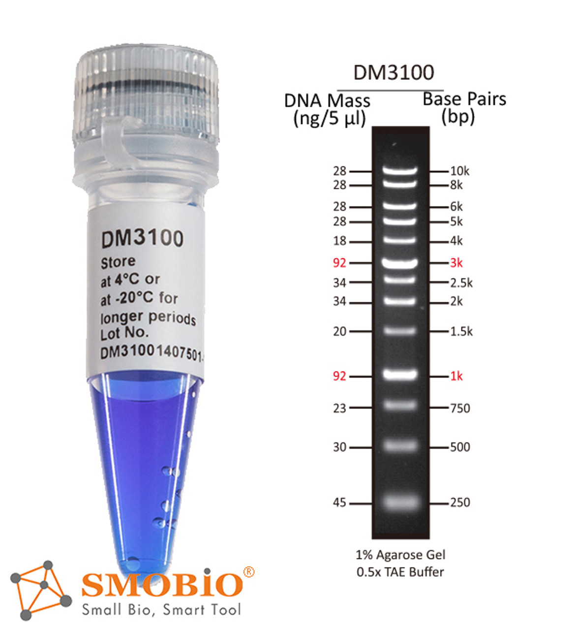
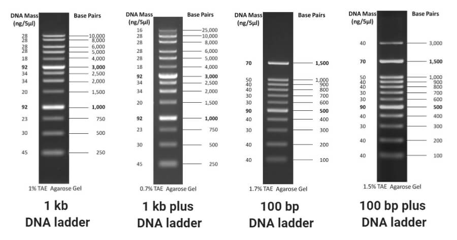
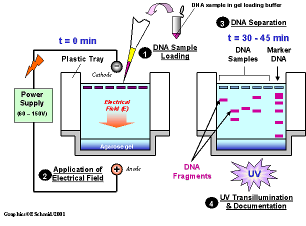
Polymerase chain reaction (PCR) (article) | Khan Academy The results of a PCR reaction are usually visualized (made visible) using gel electrophoresis. Gel electrophoresis is a technique in which fragments of DNA are pulled through a gel matrix by an electric current, and it separates DNA fragments according to size. A standard, or DNA ladder, is typically included so that the size of the fragments ... Annotating A Gel | Get Your Science On Wiki | Fandom Annotation: 1.In Inkscape import your gel file and adjust the size of your picture to fit the page out line (increase zoom if needed). 2. Add in the significant ladder measurements. (On Mark's Lab area wall or just ask Mark!) 3. Create color coded rectangles to give a background for the following text. 4. Gel electrophoresis (article) | Khan Academy Gel electrophoresis is a technique used to separate DNA fragments (or other macromolecules, such as RNA and proteins) based on their size and charge. Electrophoresis involves running a current through a gel containing the molecules of interest. Based on their size and charge, the molecules will travel through the gel in different directions or at different speeds, allowing them to be separated ...
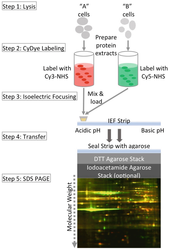
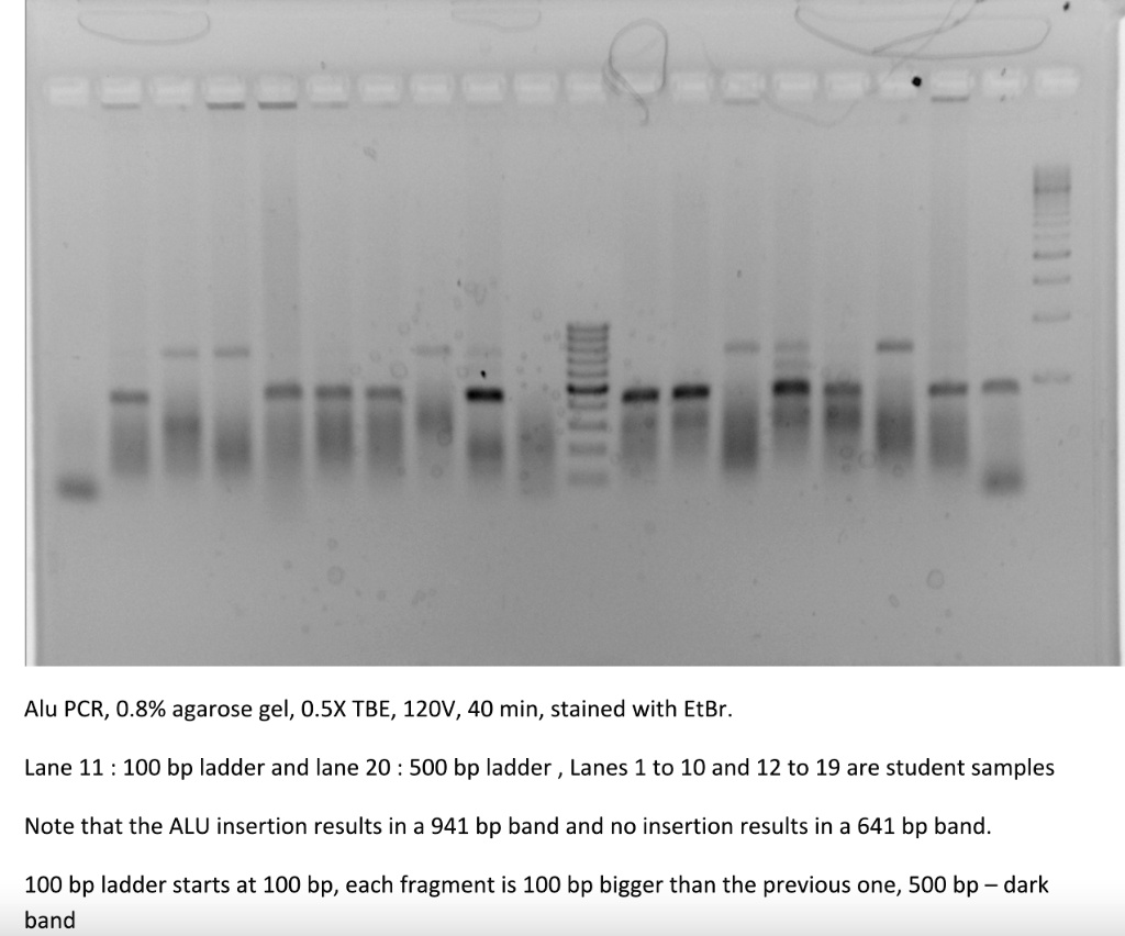
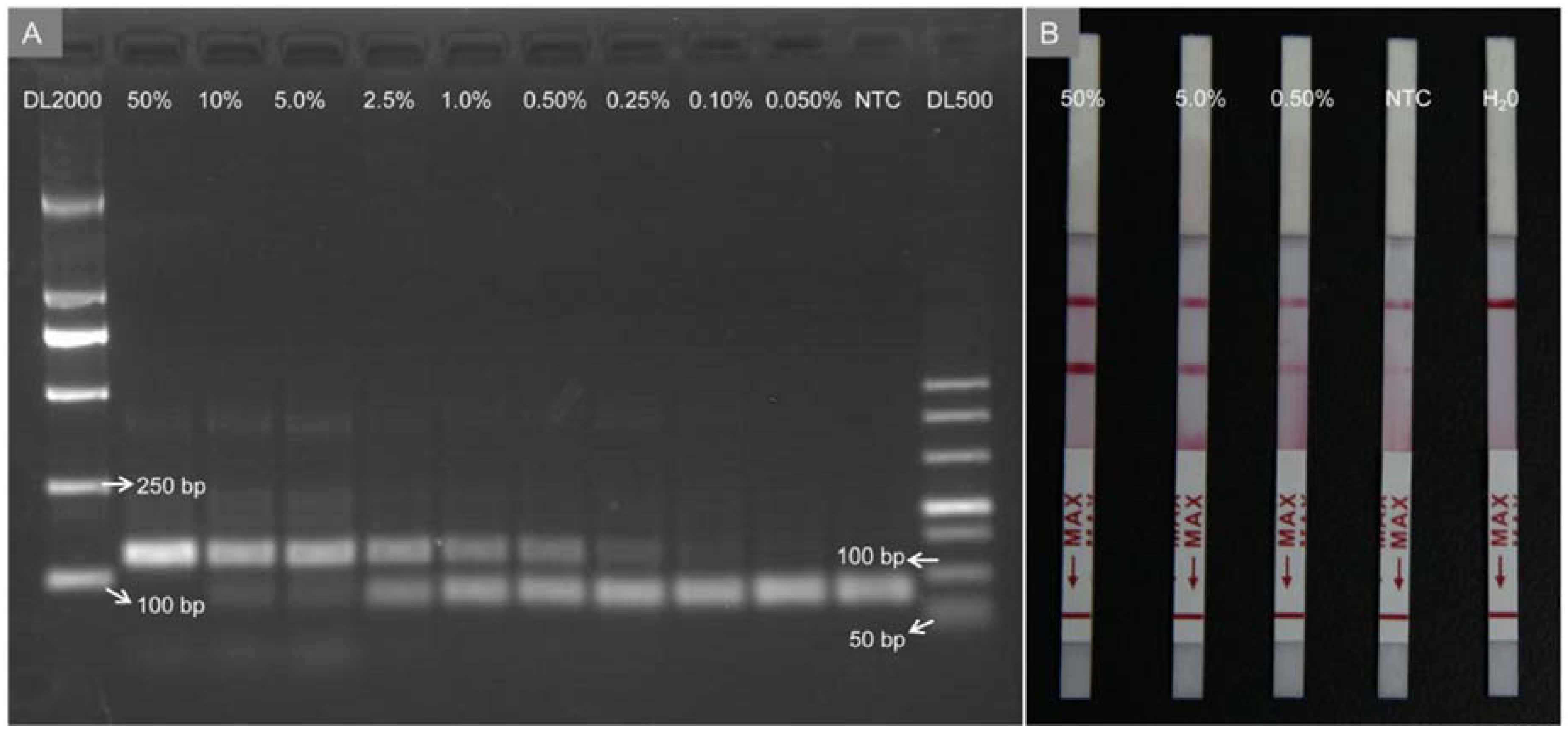
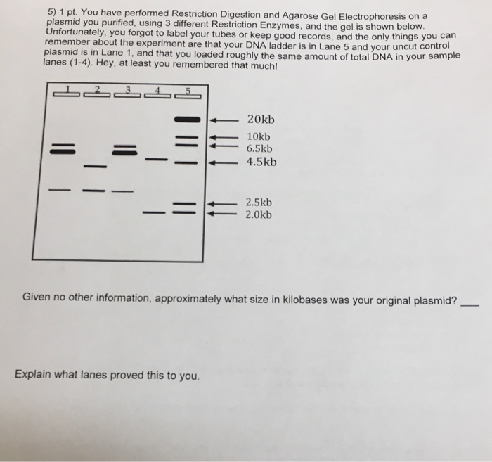
How to label gel electrophoresis images. How to Read, Interpret and Analyze Gel Electrophoresis Results? The results of PCR are run on 2% gel with a clear and known DNA ladder. Now take a look at some of the results of PCR. Image 1: PCR gel electrophoresis result image 1. The image is captured under the UV transilluminator instead of the gel doc system to show you the effect of EtBr on the gel electrophoresis results. PDF DIFFERENCE GEL ELECTROPHORESIS Imaging and Analysis of DIGE Difference gel electrophoresis (DIGE) is a relatively straightforward application of differential labeling of protein samples using fluorescent dyes. The technique, originally published by Unlu et al. (1997), uses cyanine (Cy) dyes to differentially label proteins separated by 2-D gel electrophoresis. Following labeling and electrophoresis ... How to quantify each band in gel electrophoresis? - ResearchGate Most recent answer. You can use your ladder as reference point. Measure the Rf distances for each band. Then measure the distances for thd ladder of interest. Use the logarithm on the ladder ... Part 2: Analysing and Interpreting (Agarose) Gel Electrophoresis Results The negatively charged DNA migrates towards the positive node under the influence of the current. The results of agarose electrophoresis are affected by some of the factors enlisted below, The concentration of gel. Re-use of chemicals and solutions. Unpure DNA samples.
Gel Electrophoresis Image Analysis - Thermo Fisher Scientific Gel electrophoresis analysis with E-Editor. The E-Editor 2.0 includes image editing features, such as: Image reconstruction by re-aligning and re-arranging lanes of the precast gel for easy viewing. Options to save the reconstructed image for further analysis. Image integration to group multiple images for easy comparison. GelAnalyzer Free desktop app for 1D gel electrophoresis evaluation Analyze gel images from any source. Use your digital camera, smartphone, or gel doc system to obtain images. GelAnalyzer will take care of the rest. Automatic lane and band detection. With full manual control over adding, modifying, and deleting lanes and bands. ... How to label Gel electrophoresis pictures for thesis and research ... #Gelelectrophoresis #Pictures_labelling #Research_articles @Thesis #Labelling #Pictureslabellinging_in_paint #Image_labelling #Picture_editing #cellbiology ... 3 Ways to Read Gel Electrophoresis Bands - wikiHow 1. Hold a UV light up to the gel sheet to reveal results when using a UV-based dye. With your gel sheet in front of you, find the switch on a tube of UV light to turn it on. Hold the UV light 8-16 inches (20-41 cm) away from the gel sheet. Illuminate the DNA samples with the UV light to activate the dye and read the results.
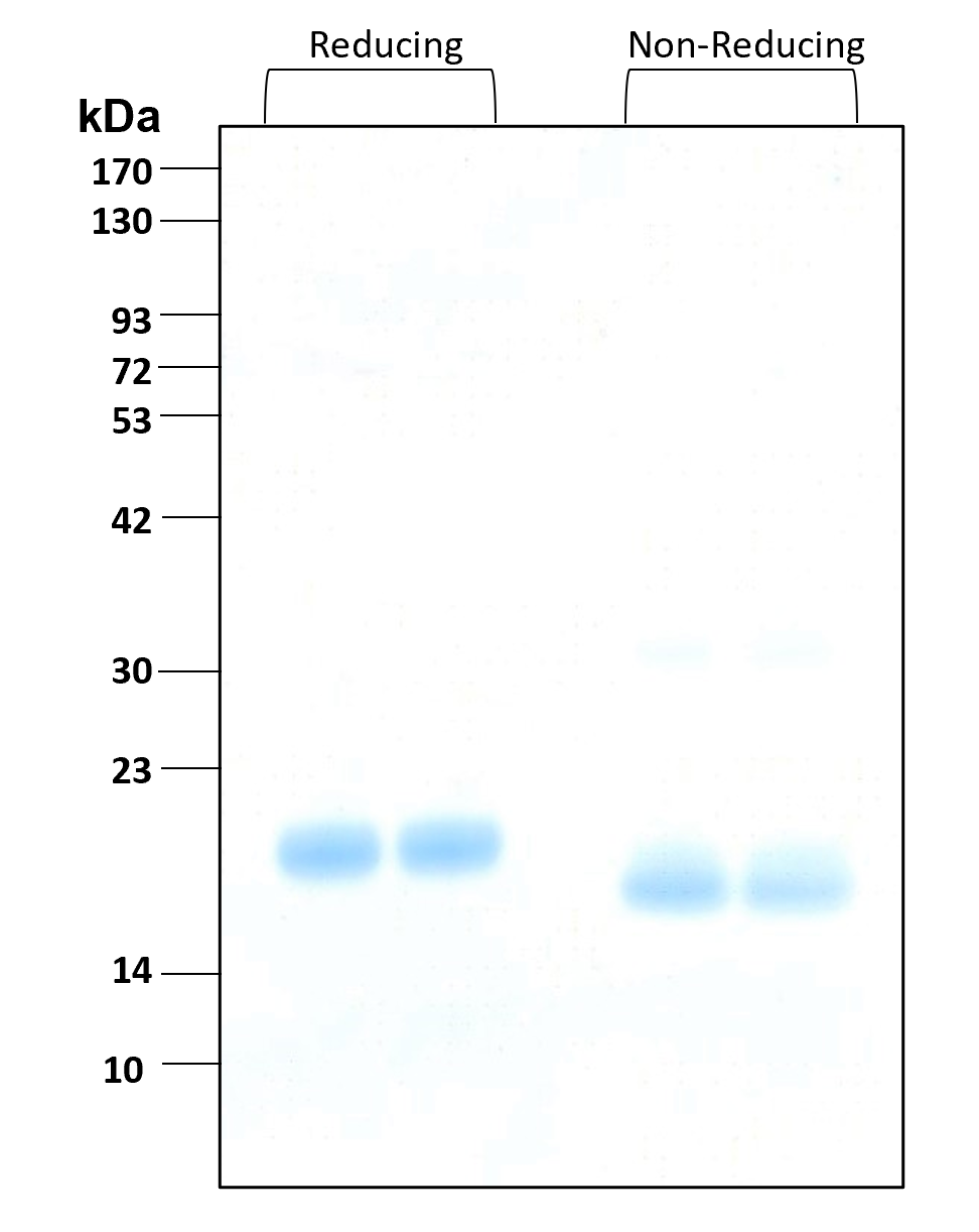
InDesign Labeling / Annotating PCR Gel Pictures - YouTube In this tutorial we will learn how to annotate Agarose Gel Pictures with Adobe InDesign CS5. I see people often labeling pictures in Photoshop and I can't re... 1.15: SDS-PAGE - Biology LibreTexts Place the lid on the vertical gel chamber. Insert the red and black wires into the correct matching colored terminals on the power supply. Plug in the power supply and turn on the power switch. Select "Constant Voltage" and then adjust the voltage to 300 volts. Press the run button. 1.12: Restriction Digest with Gel Electrophorisis Check that the gel is oriented with sample wells closest to the negative electrode (black). Check that the power cord can reach easily. Check that the gel box will not need to be moved for 30 minutes. Draw and label in your notebook how the samples will be loaded in the gel. Check whether you will be sharing the gel with another group. Texture analysis in gel electrophoresis images using an integrative ... Texture information could be used in proteomics to improve the quality of the image analysis of proteins separated on a gel. In order to evaluate the best technique to identify relevant textures ...
ImageJ for Editing & Labelling PCR Gel Image | Biotechnology This Tutorial is all about how to quickly Edit & Label PCR Gel Image Using ImageJ software. Presented by - Elvis SamuelJoin Our Telegram Channel for free Sof...
DNA sequencing (article) | Biotechnology | Khan Academy The modified image is licensed under a (CC BY-SA 3.0) license. After the reaction is done, the fragments are run through a long, thin tube containing a gel matrix in a process called capillary gel electrophoresis. Short fragments move quickly through the pores of the gel, while long fragments move more slowly. ...
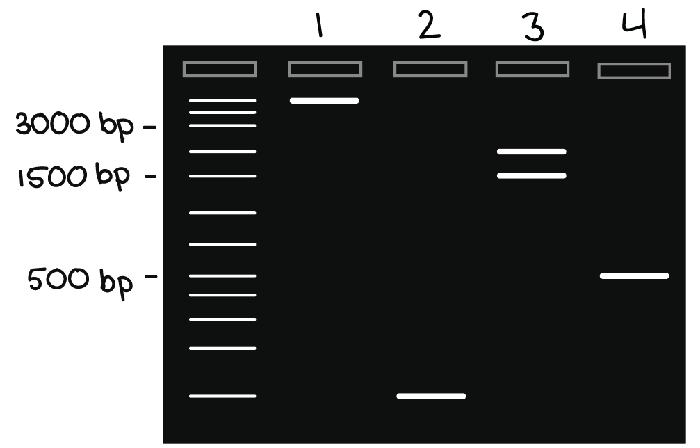
Gel electrophoresis (article) | Khan Academy Gel electrophoresis is a technique used to separate DNA fragments (or other macromolecules, such as RNA and proteins) based on their size and charge. Electrophoresis involves running a current through a gel containing the molecules of interest. Based on their size and charge, the molecules will travel through the gel in different directions or at different speeds, allowing them to be separated ...
Annotating A Gel | Get Your Science On Wiki | Fandom Annotation: 1.In Inkscape import your gel file and adjust the size of your picture to fit the page out line (increase zoom if needed). 2. Add in the significant ladder measurements. (On Mark's Lab area wall or just ask Mark!) 3. Create color coded rectangles to give a background for the following text. 4.
Polymerase chain reaction (PCR) (article) | Khan Academy The results of a PCR reaction are usually visualized (made visible) using gel electrophoresis. Gel electrophoresis is a technique in which fragments of DNA are pulled through a gel matrix by an electric current, and it separates DNA fragments according to size. A standard, or DNA ladder, is typically included so that the size of the fragments ...











Post a Comment for "40 how to label gel electrophoresis images"