43 cows eye labeled
Cow Eye Dissection & Labeling - YouTube About Press Copyright Contact us Creators Advertise Developers Terms Privacy Policy & Safety How YouTube works Test new features Press Copyright Contact us Creators ... PDF Cow Eye Dissection Guide - Central Bucks School District DISSECTION OF THE COW EYE Please make sure to wear gloves and safety glasses when you are dissecting, and make sure to clean up thoroughly after the lab. Also, the cow eyes can be rather slippery, so use caution when handling and cutting them. You will need a scalpel and forceps. 1. First, identify the most external structures of the eye.
Cow Eye for Dissection | Cow Eyeball Dissection Specimen | HST DESCRIPTION. Cow eye dissection is popular because a cow's eye is similar to the human eye! Using a scalpel, middle school and high school students can explore these preserved cow eyes to explore the amazing ways in which a cow's eye works (and thus, how a human's eye works as well)! Examine the different parts of the eye, such as the cornea at ...

Cows eye labeled
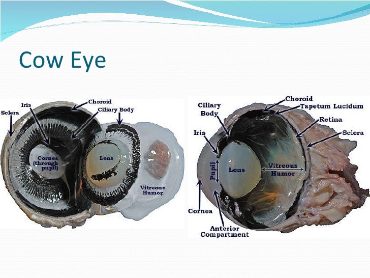
Ultrasonographic anatomy of the bovine eye - PubMed The ultrasonographic appearance of structures within the bovine eye is similar to that in other species, although the ciliary artery was frequently identified, appearing as a 0.33 +/- 0.04 cm diameter hypoechoic area. The axial length of the globe was significantly greater in Holstein Friesian cattle (3.46 +/- 0.09 cm) compared with Jersey ... Cow Eye Dissection Lab from Anatomy and Physiology The blue covering over the front of the eye is the cornea . When the cow was alive, the cornea was clear. Together with the lens, the cornea refracts light and helps the eye to focus. The cornea gives a larger contribution to the total refraction than the lens. The curvature of the cornea is fixed while that of the lens is changeable. [Solved] How to label a cow eye dissection? | Course Hero When labeling a cow eye dissection, it is also important to use clear and concise labels. Use abbreviations if necessary, but make sure that the labels are still understandable. For example, you might label the cornea as "C" and the iris as "I". In general, it is best to label the parts of the eye before you start the dissection.
Cows eye labeled. Cow Eye Dissection Parts Labeled - All About Cow Photos Solved 6 The Images Below Show A Preserved Cow S Eye One Chegg Cow Eye Dissection Google Slides Dissection Cattle Anatomy Human Eye Png Clipart Biology Blue Glow Diagram Neur 320 Art And Vision 3 Use The Pictures Below To Name Parts Of Eye That You Will Observe In Course Hero Eye Dissection Cow S Eye Dissection Diagram Cow Eye Labeling Quiz - PurposeGames.com This is an online quiz called Cow Eye Labeling. There is a printable worksheet available for download here so you can take the quiz with pen and paper. Your Skills & Rank. Total Points. 0. Get started! ... Label the parts of the digestive system 14p Image Quiz. Anterior Extrinsic(Ankle Muscles) 4p Image Quiz. Female Reproductive Labeling bc 4p ... cow eye anatomy Flashcards and Study Sets | Quizlet Cow Eye Anatomy cornea Sclera iris The clear tissue that covers the front of the eye white of the eye; maintains the shape of the eye and protects… a ring of muscle tissue that forms the colored portion of the… 14 Terms ObliviousRain PLUS Anatomy Cow/Sheep Eye Sclera Cornea Optic Nerve Gives the eye its shape and protects the inner parts. Cow Eye Dissection Guide - Google Slides Cow Eye. Use the point of a scissors or a scalpel to make an incision through the layers of the eye capsule (similar to figure 1); there are three layers from the exterior: sclera, whitish/grey, continuous with the transparent cornea, choroid, thin dark black layer and the retina, thin greyish/pink layer. Use a scissors to dissect the entire ...
Cow eye - dissection and label - SlideShare 3. Cow eye-shown with labeled Sclera The sclera (from the Greek skleros, meaning hard [1]), also known as the white of the eye, is the opaque, fibrous, protective, outer layer of the eye containing collagen and elastic fiber. 4. Cow eye-shown with lens dissected. Cow Eye Dissection Teaching Resources | Teachers Pay Teachers This is a comprehensive dissection guide of the cow eye, designed for a high school or early college Biology or Anatomy & Physiology class. The guide includes step-by-step instructions and labeled diagrams that will lead students through the external anatomy of the eye, followed by dissection of the internal structures. label the eye anatomy diagram Eye cow labeled cows enucleation unlabeled coronal section bio201 return preserved. Label the eye Labeling Parts Of the Heart Lovely Parts the Heart Proprofs Quiz we have 8 Pics about Labeling Parts Of the Heart Lovely Parts the Heart Proprofs Quiz like Label the Eye, Perch Dissection - Biology 11 Honours - Animalia Labs and also LAS Assignment ... Cow Eye Dissection & Anatomy Project | HST Learning Center Look carefully at the preserved cow eye. The most noticeable part of the eye is the large mass of gray tissue that surrounds the posterior (back) of the eye and is attached to the sclera. The second most noticeable part of the eye is the cornea, located in the anterior (front) part of the eye.
PDF COW'S EYE dissection - Exploratorium get cows' eyes: You can order cows' eyes at a butcher shop or purchase them directly from a slaughterhouse. Try to get eyes with the muscles and fat still attached. If possible pick up the cows' eyes the day of the dissection; eyes are easier to cut when they are fresh. This diagram shows the parts of the eye. Dissecting An Eyeball - Krieger Science Cow eyes are also readily available, as a byproduct of beef production, and can be purchased inexpensively from science supply companies, such as Carolina Biological Supply. Such eyeballs are normally stored in preservative, which has the unfortunate side effect of making the cornea and lens cloudy, but the interior of the eyeball is still ... PDF Name: Dissection 101: Cow Eye human eye _____ _____ Draw and label the cow eye. Cornea Optic nerve Vitreous humor Retina Optic disc (blind spot) Choroid Tapetum lucidum Sclera Aqueous humor Suspensory ligaments Lens Ciliary body Pupil Iris Provided by Dissection 101: Cow Eye Cow Eye Dissection - The Biology Corner 1. Examine the outside of the eye. You should be able to find the sclera, or the whites of the eye. This tough, outer covering of the eyeball has fat and muscle attached to it 2. Locate the covering over the front of the eye, the cornea. When the cow was alive, the cornea was clear. In your cow's eye, the cornea may be cloudy or blue in color. 2.
Vintage Popcorn Tin 1977 Colorado Country Gold Snack | Etsy | Popcorn ...
Cow's Eye Dissection - Eye diagram - Exploratorium A muscle that controls how much light enters the eye. It is suspended between the A cow's iris is brown. many colors, including brown, blue, green, and gray. A clear fluid that helps the cornea keep its rounded shape. The pupil is the dark circle in the center of your iris. It's a hole that Your pupil is round.
Cow
Cow Eye Dissection | Carolina.com Students explore the external and internal anatomy, learning how structures work together to create images from incoming light. A preserved cow eye dissection can be carried out in 1-2 class periods and only requires basic dissecting instruments. Explore the internal and external anatomy of the cow eye using the procedural steps below.

Cow Eye Dissection Parts Labeled - All About Cow Photos
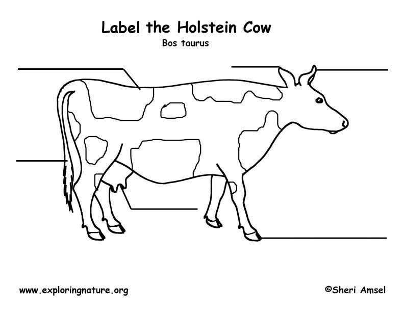
Cow Anatomy - External Body Parts and Internal Organs with Labeled ... The external body parts from the head region of a cow - in this head region, you might identify the mouth, lip, cheek, chin, muzzle, forehead, poll, ear, eye, nostril, and other. Different parts from the neck region of a cow - here, you will find the neck crest, dewlap, brisket, and jugular groove.

Rocker O Ranch - Home | Facebook
PDF Cow Eye Dissection Lab - Home Science Tools This cow eye dissection kit comes with everything you need to conduct a lab examination. Safety Guidelines • Work in a place separate from eating and food preparation areas. • Use disposable latex gloves or nitrile gloves during the dissection and cleanup. • Use only dissection tools provided.

Cow Eye Dissection Vitreous Humor | BlageusDown
Cow Eye Dissection Kit for Kids Animal Anatomy Labs | HST Product Description. Use this cow eye dissection kit to get an inside view of how the eye works! A cow eye dissection is a memorable way for elementary, middle school, and high school students to learn about anatomy and life science. This complete dissection kit provides an exceptional home or classroom experience for curious learners of all ages!

Nutmeg, our La Mancha goat | Homesteady | Pinterest | Goats, Animal and ...
Cow Eye Dissection & Parts of the Eye Diagram | Quizlet cornea Clear, outer layer of the front of the eye. sclera White, outermost layer of the eye. Helps maintain shape and gives attachment to muscles. photoreceptors The cells in the retina that respond to light (rods and cones) rods Photoreceptor cells in the eye that detect black, white, and gray cones Photoreceptor cells in the eye that detect color
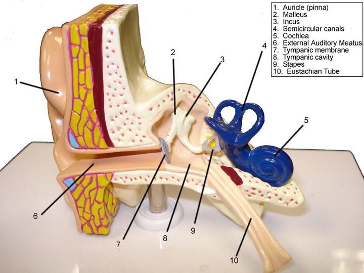
Lab Guide: Ear, Eye, Brain
Cow Eye Parts Labeled - All About Cow Photos Cow Eye Labeled Clipart Best Development Anatomy And Physiology Of The Eye Word Perspective Es From Latin Per Through Specere Look Anatomy Of The Eye Sheep Eye Dissection Lab Lab 15 Cow Eye Dissection Flashcards Quizlet Cow S Eye Diagram Quiz Ppt Cow Eye Dissection Powerpoint Ation Id 3482425 Cow Eye Dissection Anatomy Hst Learning Center

HBS - Labeling the Cow Eye Quiz - Quizizz Play this game to review undefined. Label the aqueous humor. Preview this quiz on Quizizz. Label the aqueous humor. HBS - Labeling the Cow Eye DRAFT. 10th - 12th grade. 112 times. 86% average accuracy. 7 months ago. vcaudill. 1. Save. Edit. Edit. HBS - Labeling the Cow Eye DRAFT. 7 months ago. by vcaudill. Played 112 times. 1. 10th - 12th grade ...
Eye anatomy
PDF Table 8.1: External Anatomy of The Cow Eye Feature Description TABLE 8.2 CONTINUED: INTERNAL ANATOMY OF THE COW EYE FEATURE DESCRIPTION Retina Innermost layer of the eye; only in posterior cavity; delicate, thin, cream colored sheet of tissue Optic disc A single point of attachment of the retina - to the optic nerve (also called the blind spot) Choroid Middle layer of the eye, posterior portion;
Post a Comment for "43 cows eye labeled"